Category: Science
-
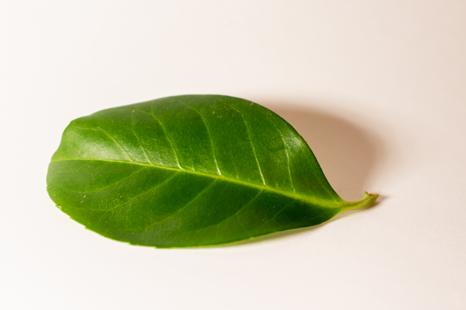
Laurus nobilis
We can see them everywhere as garden separation but how Laurel (Laurus nobilis) looks like under a Microscope? ” order_by=”sortorder” order_direction=”ASC” returns=”included” maximum_entity_count=”500″]
-
Milk under microscope (100x with oil)
What milk looks like under a microscope? You can find here a video of Milk with a 100x (with oil) lens.
-
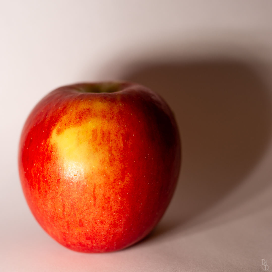
Play a song for me AppleJazz, AppleJazz
Developed in New Zealand, It was launched commercially in April 2004 (References). But how this Apple Looks like inside? Let’s start with the skin: ” order_by=”sortorder” order_direction=”ASC” returns=”included” maximum_entity_count=”500″] What about the flesh? ” order_by=”sortorder” order_direction=”ASC” returns=”included” maximum_entity_count=”500″] Why do apple slices turn brown ? When an apple is cut (or bruised), oxygen is introduced…
-
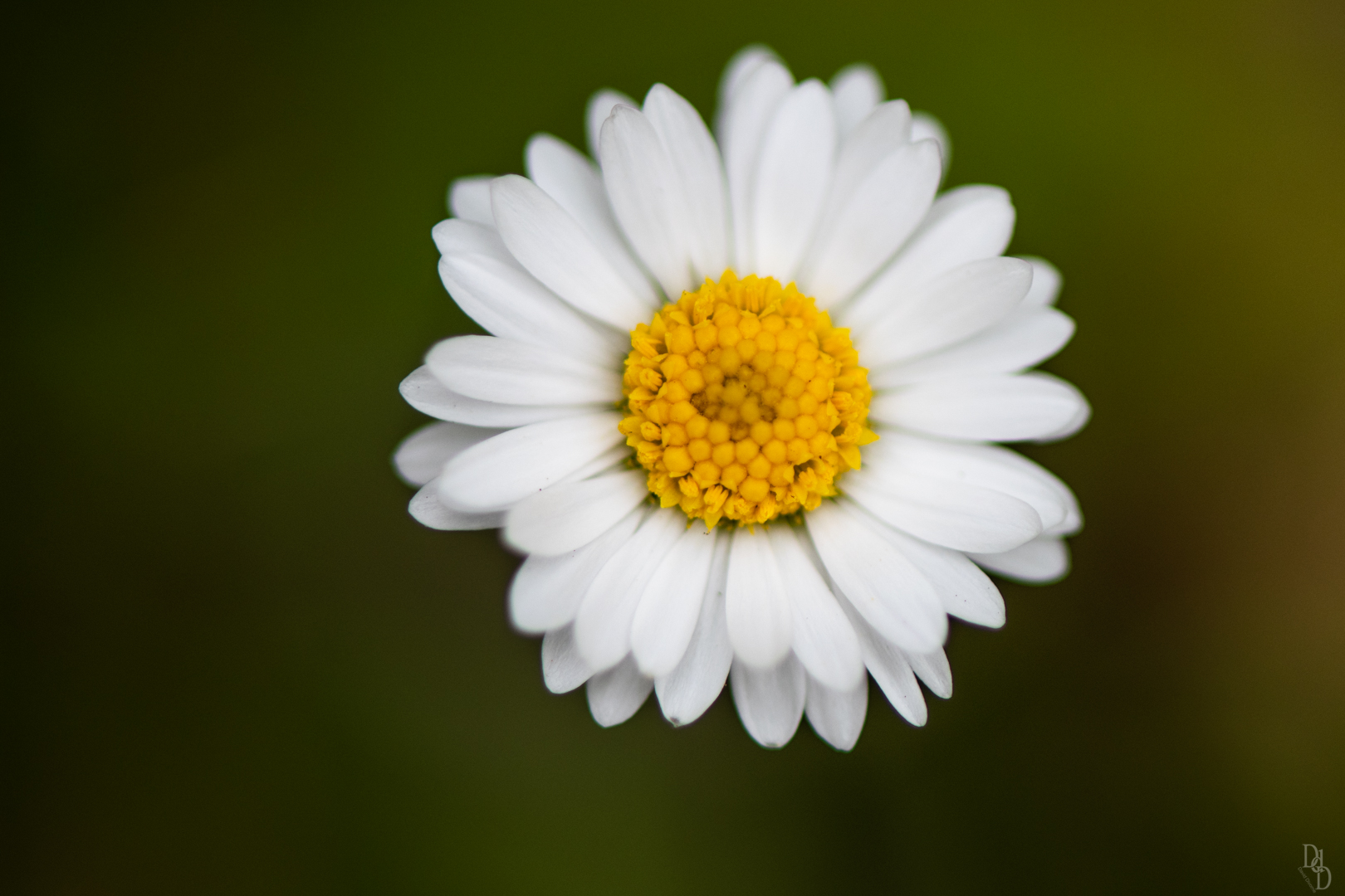
Daisy, he loves me… he loves me not
The Petal ” order_by=”sortorder” order_direction=”ASC” returns=”included” maximum_entity_count=”500″] The Pistil ” order_by=”sortorder” order_direction=”ASC” returns=”included” maximum_entity_count=”500″]
-
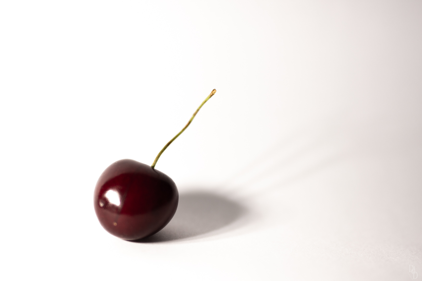
Cherry, Cherry
Time for a Cherry to be analysed Cherry skin. As for all other fruits, we can see that the skin cells are smaller but denser ” order_by=”sortorder” order_direction=”ASC” returns=”included” maximum_entity_count=”500″] Cherry Flesh ” order_by=”sortorder” order_direction=”ASC” returns=”included” maximum_entity_count=”500″]
-
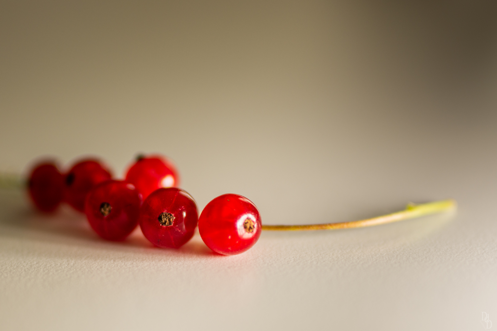
When redcurrant plays the artists
Redcurrant season is arrived! Personnally, I’m a fan of this berry as jam or pie (thanks to my wife)! But how looks like this berry under a Microscope? Let’s take a look by cutting a fragment on the slide. What about the skin? We can easily realise that the skin has an higher density…
-

Watermelon under Microscope
If you wonder what a watermelon looks like under a microscope, you are in the correct page! You can see here, the different size from the lens 4x to 100x (1000x with oil). So from to original scale: 4x 10x 40x 100x What makes the watermelon red? A pigment called Lycopene…
Recent Posts
- View on the Moming Glacier
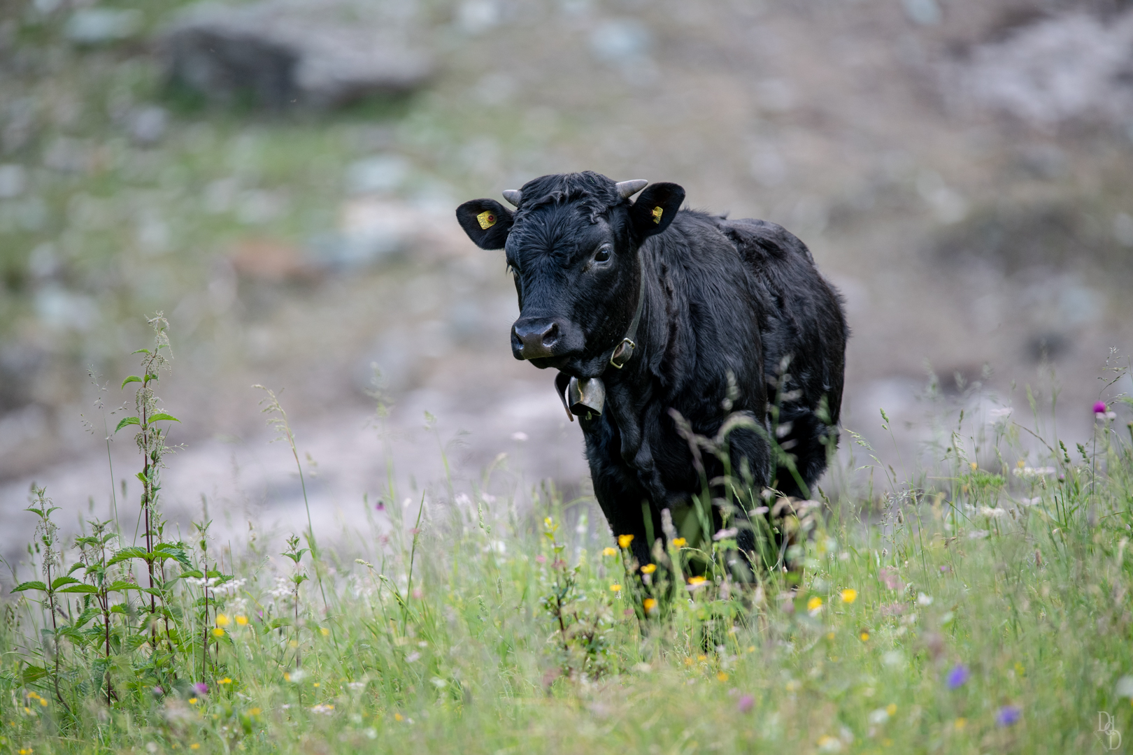
- Val d’Anniviers, Juillet 2018
- Laurus nobilis
- Milk under microscope (100x with oil)
- Play a song for me AppleJazz, AppleJazz